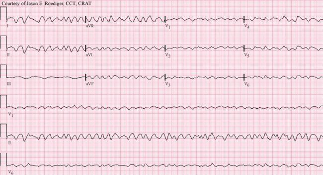
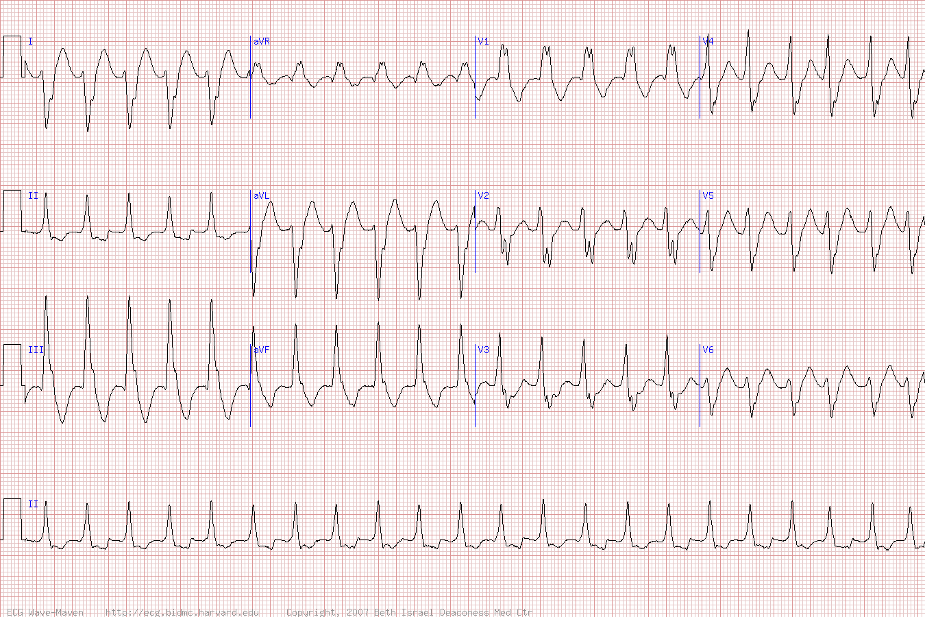
V Tach Ecg

Three or more ventricular beats with a maximal duration of 30 seconds.
V tach ecg. You will then follow the acls algorithm. Although there is a broad complex tachycardia hr 100 qrs 120 the appearance in v1 is more suggestive of svt with aberrancy given that the the complexes are not that broad 160 ms and the right rabbit ear is taller than the left. A vt of more than 30 seconds duration or less if treated by electrocardioversion within 30 seconds. This ecg is a difficult one.
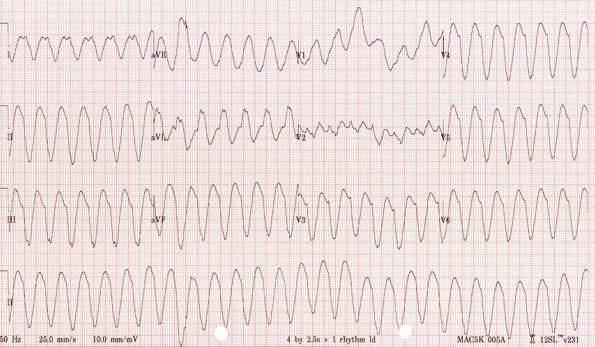
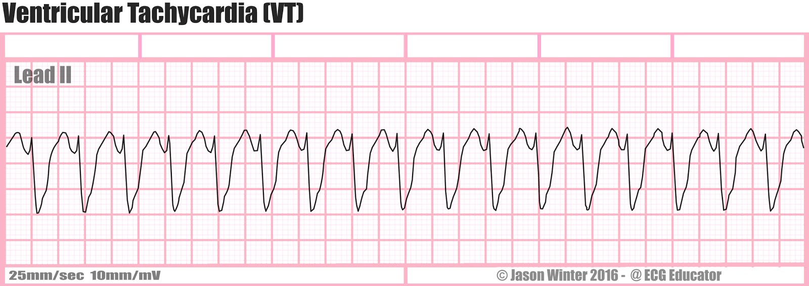
This is a very serious arrhythmia as it may lead to life threatening conditions such as ventricular fibrillation asystole and sudden death. Although there is a broad complex tachycardia hr 100 qrs 120 the appearance in v1 is more suggestive of svt with aberrancy given that the the complexes are not that broad 160 ms and the right rabbit ear is taller than the left. Ventricular tachycardia rate approximately 220 beats per minute during this rhythm she was awake protecting her airway with pallor diaphoresis and cool extremities. In monomorphic ventricular tachycardia the shape of each heart beat on the ecg looks the same because the impulse is either being generated from increased automaticity of a single point in either the left or the right ventricle or due to a reentry circuit within the ventricle.
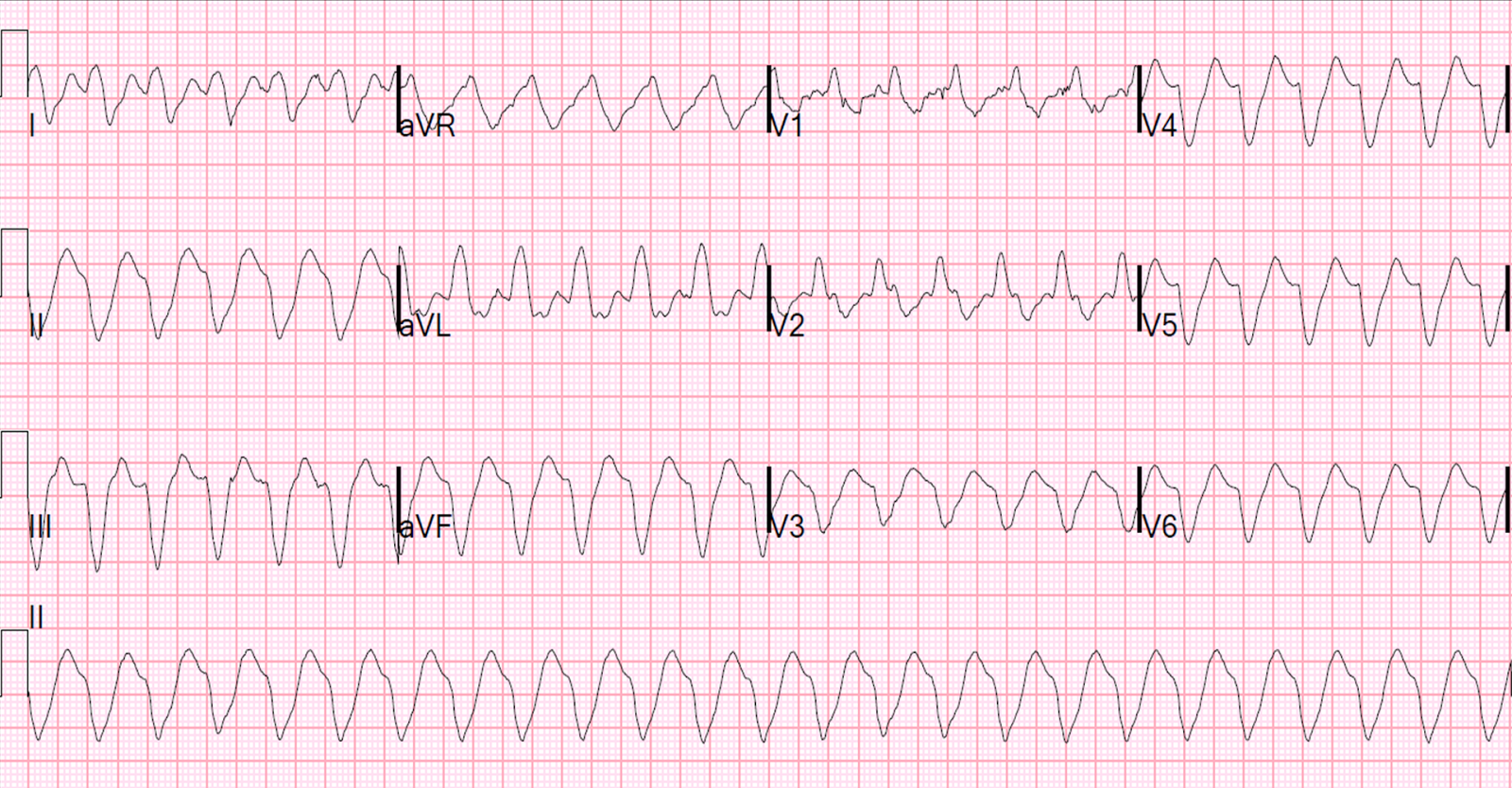
Ventricular tachycardia can be catechorized as follows. The ecg shown here is of a patient in v tach. You will need multiple nurses in the room as well as at least one provider preferably an attending doctor. There are several strong signs that this is v tach including the wide qrs complexes lack of associated p waves backward axis also called extreme right axis deviation leads ii iii and avf are all negative and avr is positive and v6 is negative.
Ventricular tachycardia vt is a cardiac arrhythmia characterized by a pulse rate of more than 100 beats per minute with at least three irregular heartbeats in a row.
